藻類の画像集 
 不等毛植物門
 
--------------------------

珪藻綱 
  (Bacillariophyceae)

 
中心型珪藻 
 タルケイソウ
   Melosira )
 ステファノピクシス
   (Stephanopyxis )
 アウラコセイラ
   Aulacoseira)
 パラリア
   (Paralia )
 リゾソレニア
   (Rhizosolenia )
 ウロコケイソウ
   (Urosolenia )
 コアミケイソウ
   Coscinodiscus )
 アクティノキクラス
   (Actinocyclus )
 アクティノプティカス
   (Actinoptychus)
 アステロムファルス
   (Asteromphalus )
 クモノスケイソウ
   Arachnoidiscus )
 キートケロス
   (Chaetoceros )
 ストレプトセカ
   (Streptotheca)
 ビドルフィア
   (Biddulphia )
 トリセラチウム
   (Triceratium )
 タラシオシラ
   Thalassiosira )
 シオノディスカス
   (Shionodiscus)
 ヒメマルケイソウ
   (Cyclotella )
 スケレトネマ
   (Skeletonema )
 ディチルム
   (Ditylum)

Fragilaria亜綱 
 フラギラリア
   (Fragilaria)
 ホシガタケイソウ
   (Asterionella)
 アステリオネロプシス
   (Asterionellopsis)
 ディアトマ
   Diatoma )
 メリディオン
   (Meridion)
 シネドラ
   (Synedra)
 ヌサガタケイソウ
   (Tabellaria )
 リコモフォラ
   (Licmophora )
 ラブドネマ
   (Rhabdonema )

羽状縦溝型珪藻 
 クチビルケイソウ
   (Cymbella )
 エンシオネマ
   Encyonema )
 クサビケイソウ
   (Gomphonema )
 ゴンフォネイス
   (Gomphoneis)
 アクナンテス
   (Achnanthes )
 ココネイス
   Cocconeis )
 アンフォラ
   Amphora )
 フナガタケイソウ
   Navicula )
 トラキネイス
   (Trachyneis )
 パルリベルス
   (Parlibellus )
 ルチコラ
   (Luticola )
 ネイディウム
   Neidium )
 フルスツリア
   (Frustulia)
 セラフォラ
   (Sellaphora)
 ハネケイソウ
   Pinnularia )
 カロネイス
   (Caloneis )
 マユケイソウ
   Diploneis )
 エスガタケイソウ
   (Gyrosigma )
 メガネケイソウ
   Pleurosigma
 スタウロネイス
   Stauroneis )
 クラチクラ
   Craticula )
 ヒッポドンタ
   (Hippodonta )
 イカダケイソウ
   Bacillaria )
 ササノハケイソウ
   Nitzschia )
 トリブリオネラ
   (Tryblionella )
 プサムディクチオン
   (Psammodictyon )
 ロパロディア
   Rhopalodia )
 スリレラ
   Surirella )
 ハダナミケイソウ
   (Cymatopleura )
 エントモネイス
   (Entomoneis )

 ユーノティア
   (Eunotia


--------------------------
黄金色藻綱 
  (Chrysophyceae)
ラフィド藻綱 
  (Raphidophyceae)
黄緑色藻綱 
  (Xanthophyceae)
ディクティオカ藻類 
  (Dictyochophyceae)

褐藻綱 
  (Phaeophyceae)

--------------------------

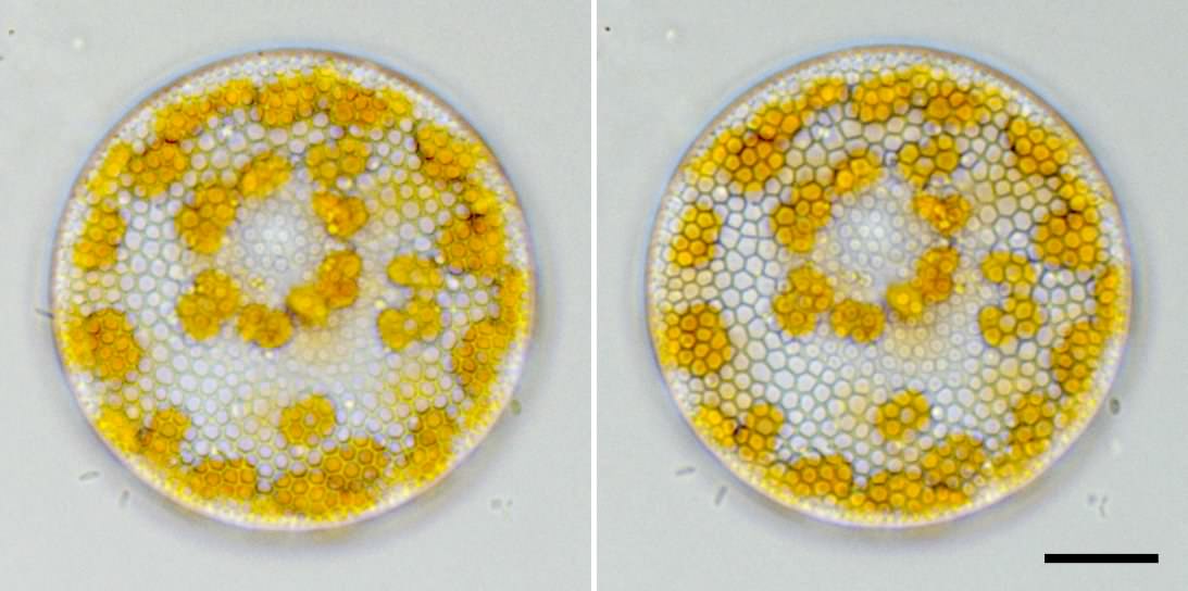
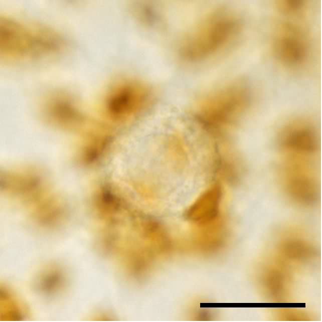
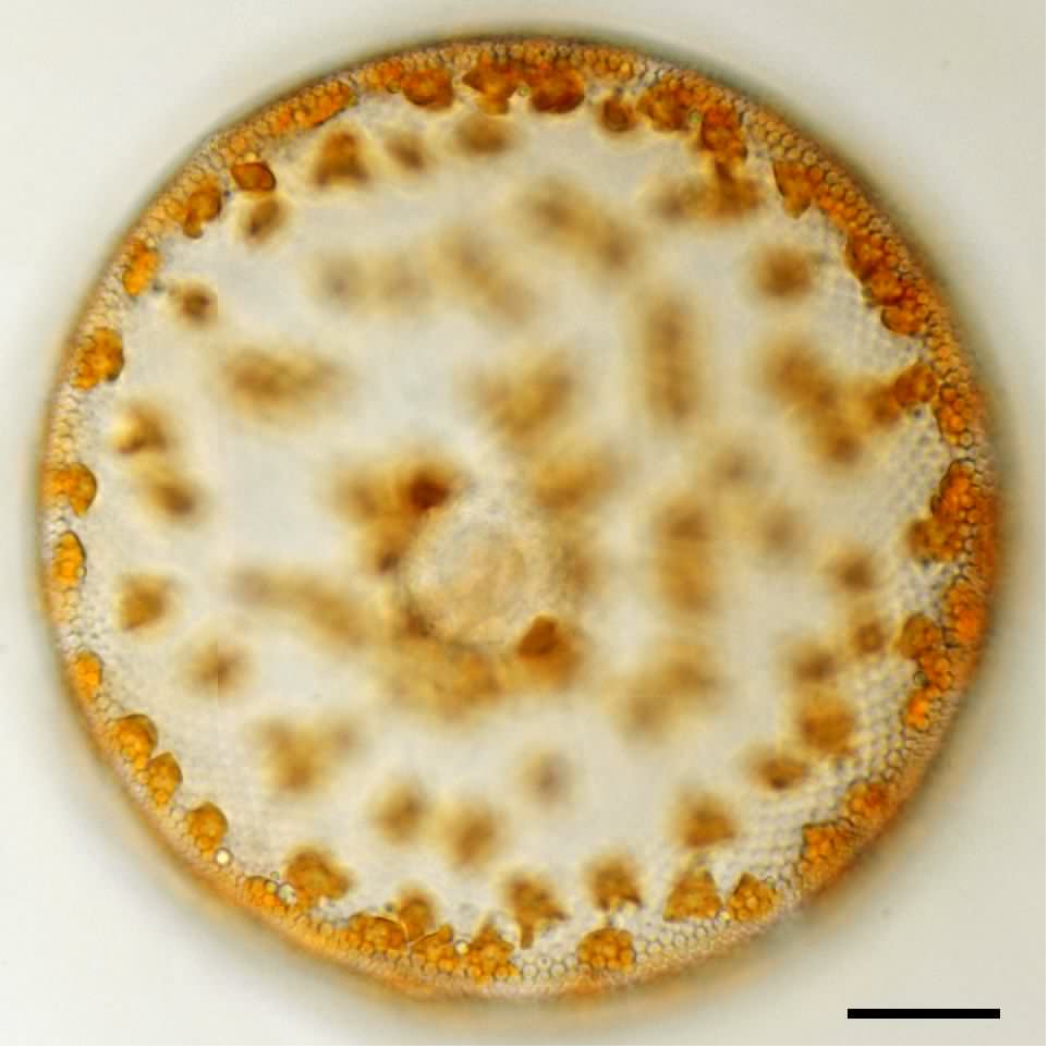
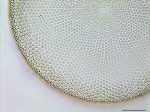
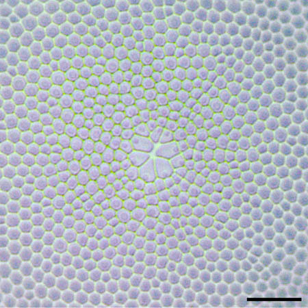
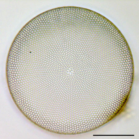
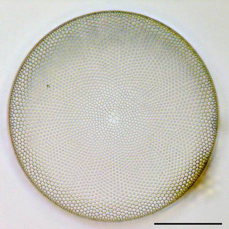
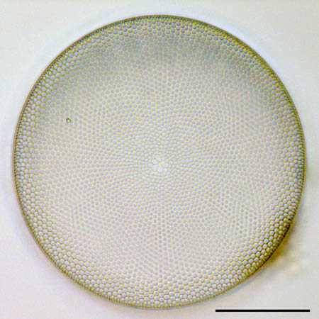
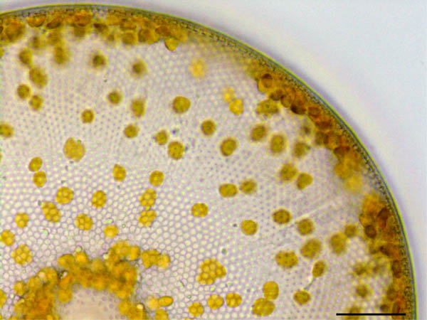
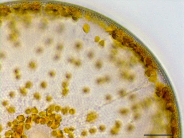
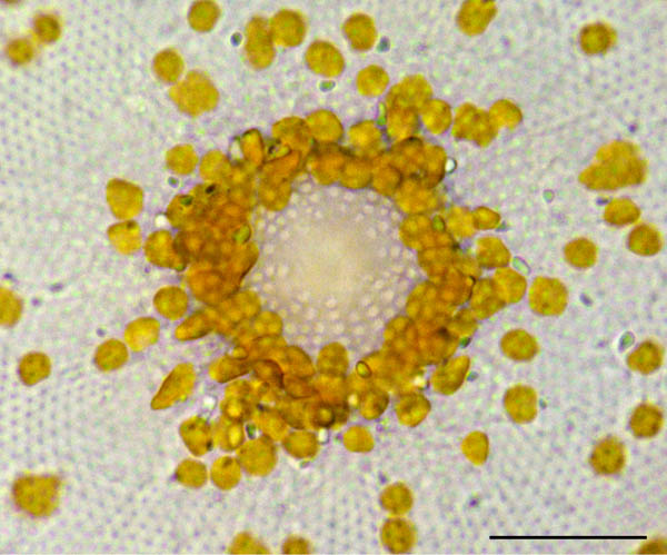
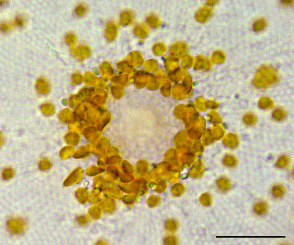
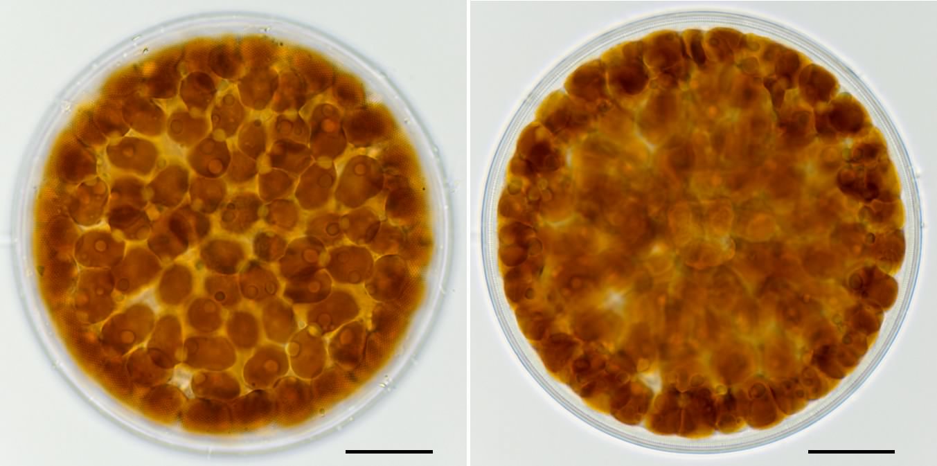
分類  :コアミケイソウ (Coscinodiscus sp.)
倍率  :1000倍 
採取  :海岸(兵庫県 大倉海岸) 
 
時期  :2008年12月採取 表面海水温13.9℃ 


 胞紋は繊細です。密度は中心域付近で15個/10μmです。


写真10a,10b

 
殻にフォーカスを合わせた画像です。
 胞紋が繊細なために蜂の巣状の構造が見えません。


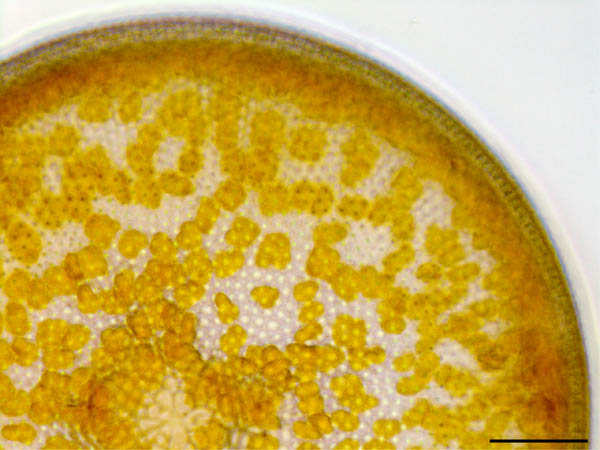
写真10c,10d
 
 
細胞内部にフォーカスを合わせた画像です。
 不定形の色素体を観察できます。
 色素体は高密度です。


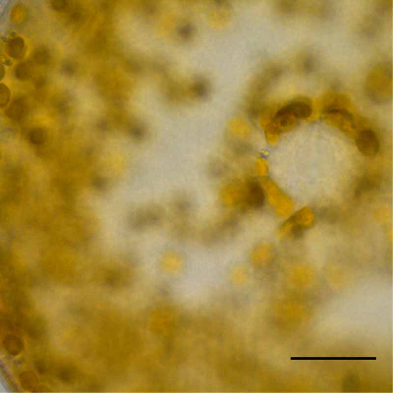
写真10e
 
 
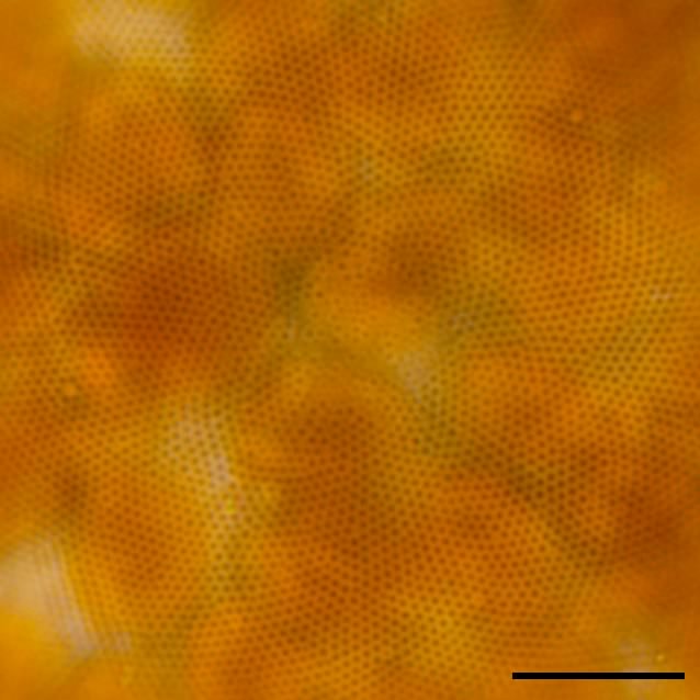
被殻の中央付近を拡大した画像です。
 胞紋の密度は15個/10μm程度です。
 中心域にはやや大型の胞紋がまばらに存在しています。
 胞紋は編心的配列をしています。


写真10f
 
 
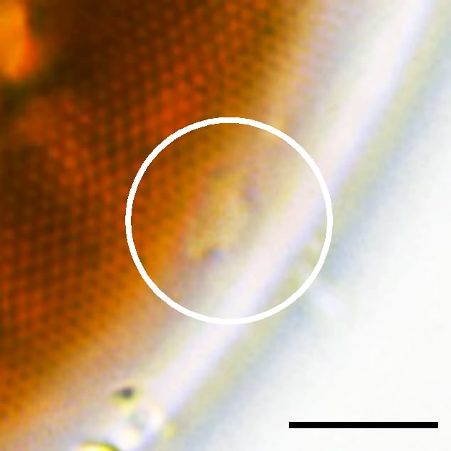
大きな唇状突起を観察できました。
写真10f
写真10e
写真10d
写真10c
写真10b
写真10a
5μm
10μm
20μm
20μm
20μm
20μm
コアミケイソウ (Coscinodiscus sp.)の画像
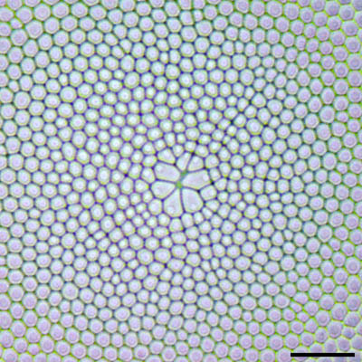
分類  :コアミケイソウ (Coscinodiscus sp.)
倍率  :1000倍 
採取  :海岸(兵庫県 須磨) 
 
時期  :2008年7月採取 表面海水温29.0℃ 


瀬戸内海で採取した小型のコアミケイソウです。
 タラシオシラ(Thalassiosira属)に似ていますが、
 被殻の内側の微細構造に違いがあります。
 タラシオシラは唇状突起と有機突起を持ちますが、
 コアミケイソウは唇状突起だけを持ちます。

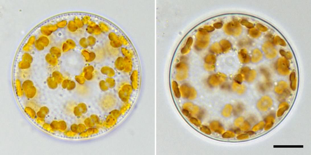
写真08a,08b
 
 被殻にフォーカスを合わせています。
 蜂の巣状の胞紋が放射状に配列しています。
 細胞の中央部(中心域)には胞紋がありません。
写真08c,08d
 
 細胞内部にフォーカスを合わせています。
 ひょうたん型の色素体と、核(矢印)があります。
写真08d
写真08c
写真08b
写真08a
10μm
10μm
コアミケイソウ (Coscinodiscus sp.)の画像
写真07a
写真07b
写真07c
写真07d
20μm
20μm
20μm
20μm
コアミケイソウ (Coscinodiscus sp.)の画像
写真06a
写真06b
50μm
50μm
コアミケイソウ (Coscinodiscus sp.)の画像
写真04a
写真04b
写真04c
写真04d
50μm
50μm
50μm
50μm
コアミケイソウ (Coscinodiscus sp.)の画像
分類  :コアミケイソウ (Coscinodiscus sp.)
倍率  :1000倍 
採取  :海岸(神奈川県 三浦半島) 
 
時期  :2014年11月採取 


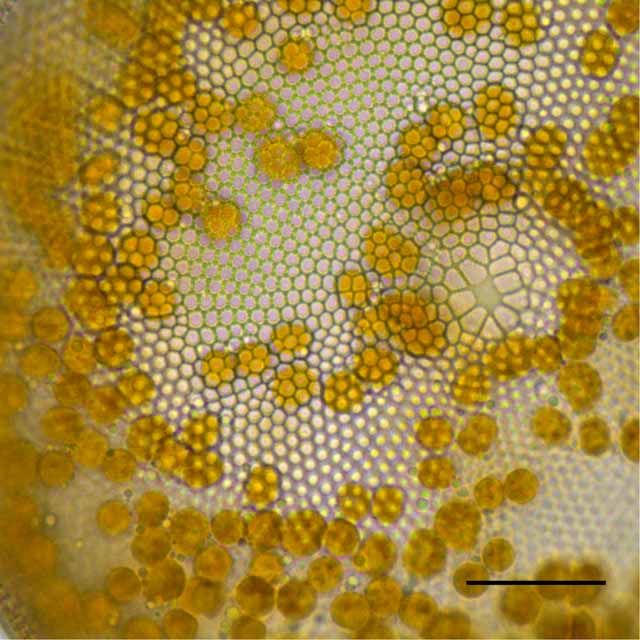
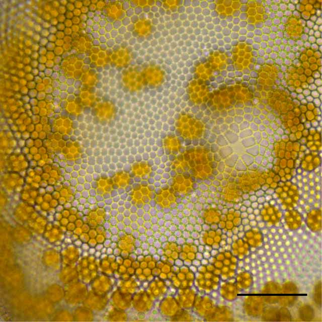
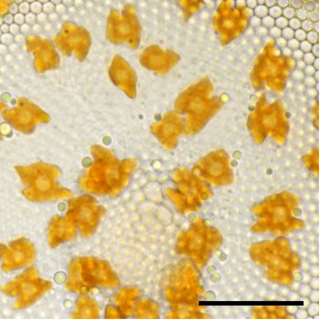
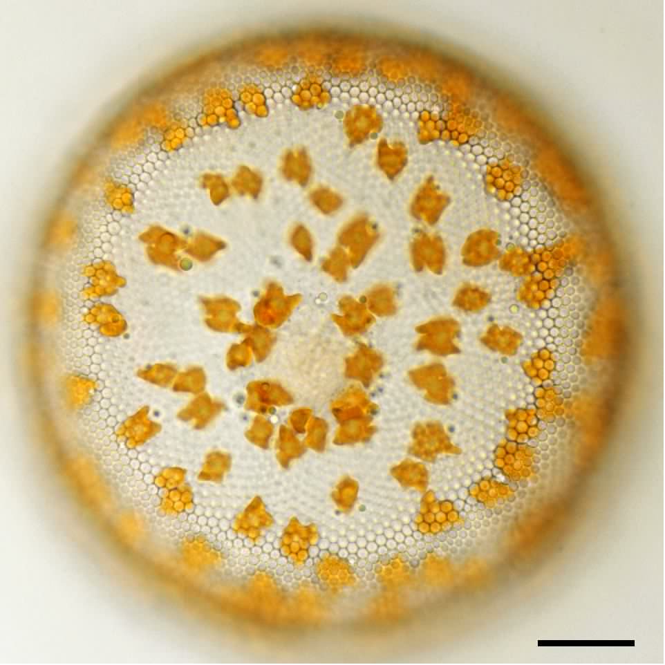
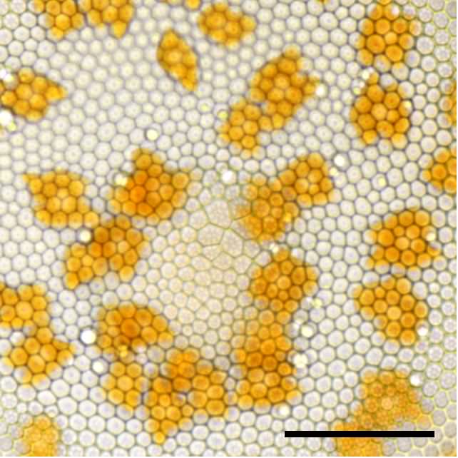
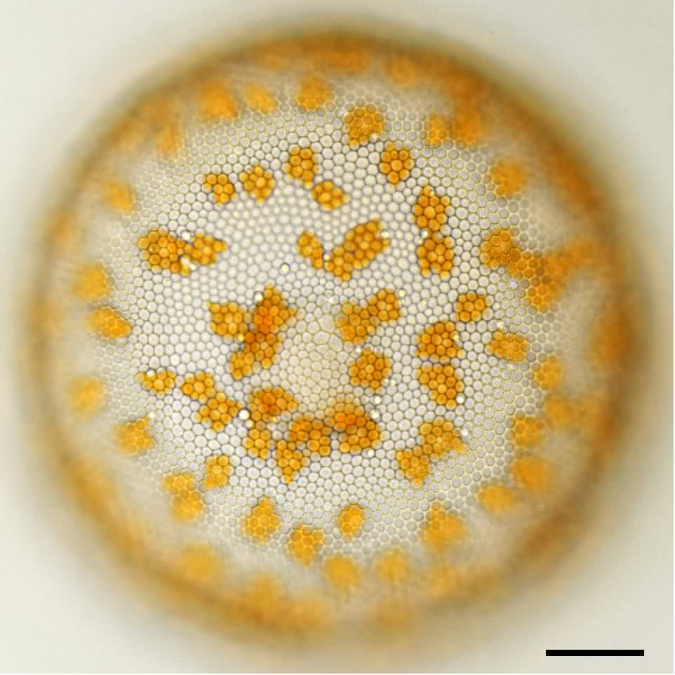
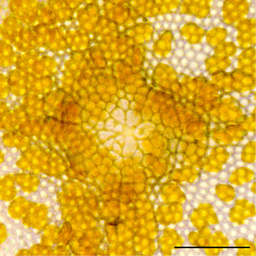
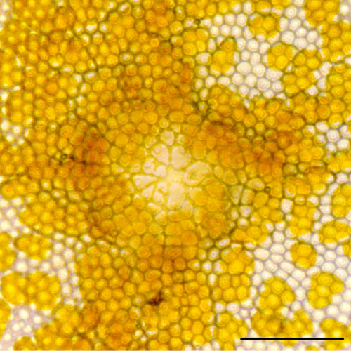
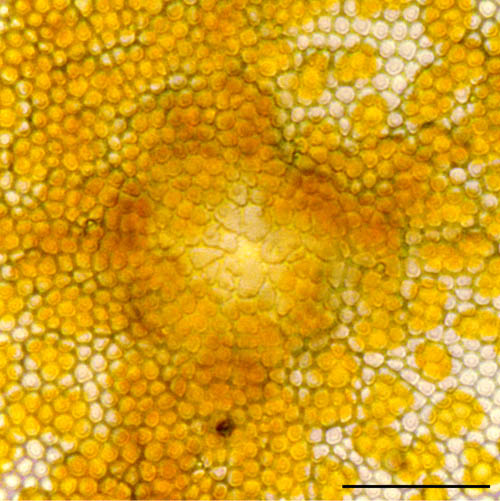
写真07a〜写真07dは写真06の中心域の拡大画像で、
 フォーカスを変えて撮影しました。
 写真07aと写真07bは殻の胞紋にフォーカスを合わせた画像です。
 写真右よりに中心域があります。
 写真07cは細胞内部の色素体にフォーカスを合わせた画像です。
 円盤状の色素体を観察できます。
 写真07dは細胞中心部の核にフォーカスを合わせた画像です。
 中心域の位置に核があります。
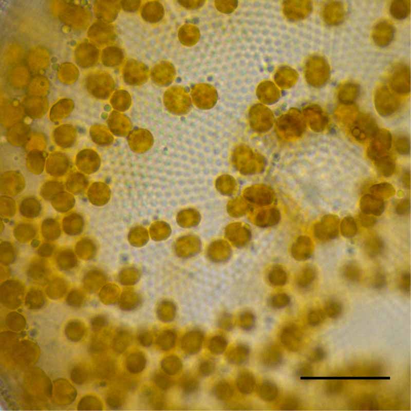
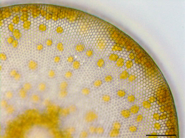
分類  :コアミケイソウ (Coscinodiscus sp.)
倍率  :500倍 
採取  :海岸(神奈川県 三浦半島) 
 
時期  :2014年11月採取 


 写真03と同時に採取した個体です。

 コアミケイソウ
(Coscinodiscus)は大きいので
 見つけやすいのですが、
 カバーガラスを被せるときに壊れやすく、
 慣れないとプレパラート作りに苦労します。
 海藻の破片を一緒に挟み込むと破壊防止になります。
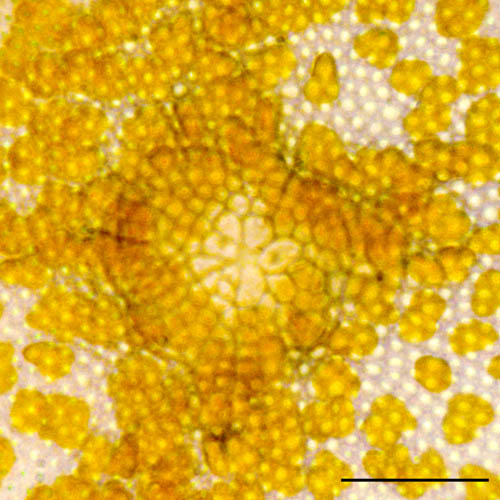
分類  :コアミケイソウ (Coscinodiscus sp.)
倍率  :1000倍 
採取  :海岸(神奈川県 三浦半島) 
 
時期  :2014年11月採取 


 写真05a〜写真05bは写真04の中心域を拡大した画像で、
 フォーカスを変えて撮影しました。
 中心域には胞紋がない領域があります。
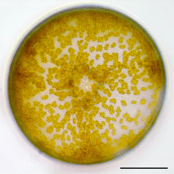
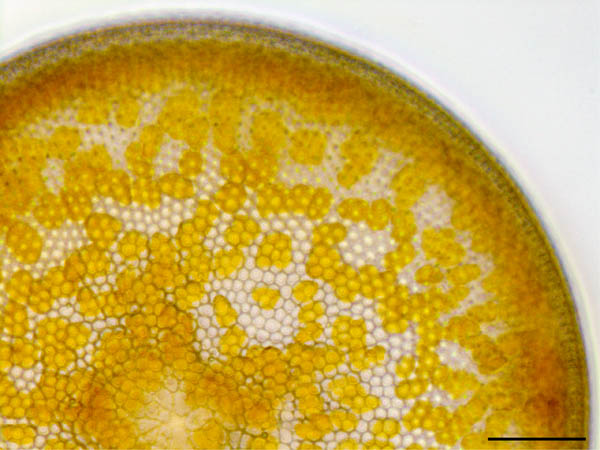
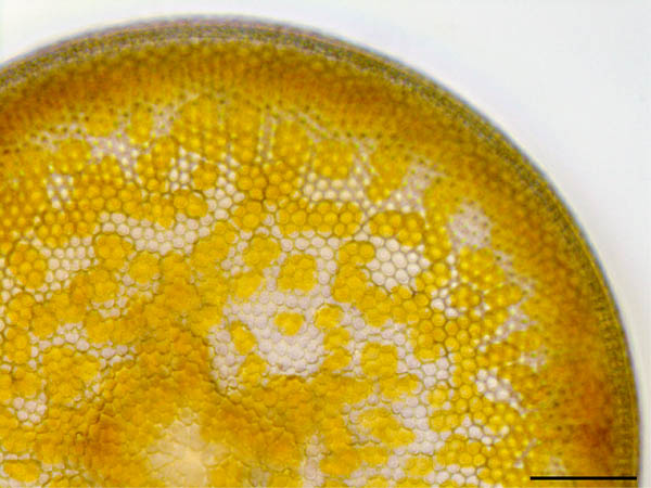
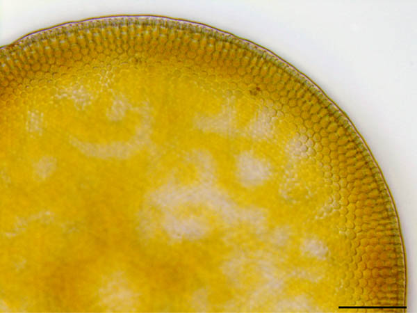
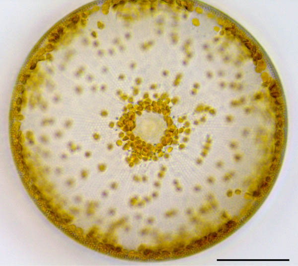
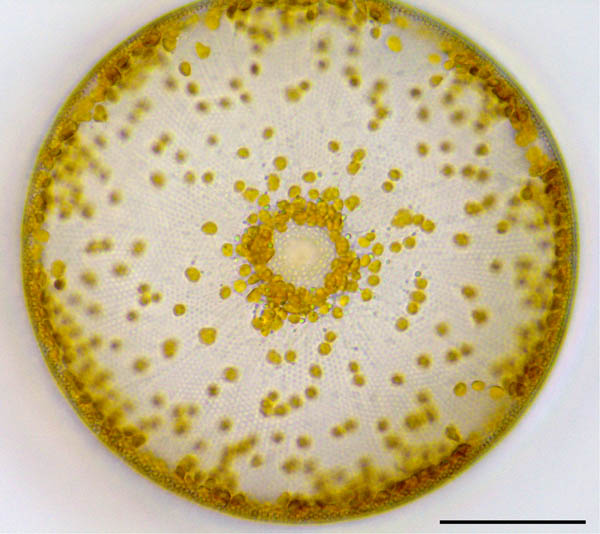
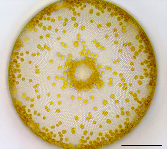
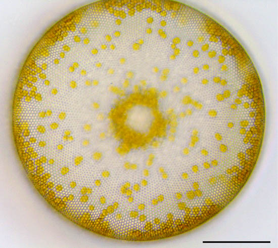
分類  :コアミケイソウ (Coscinodiscus sp.)
倍率  :500倍 
採取  :海岸(神奈川県 三浦半島) 
 
時期  :2014年11月採取 


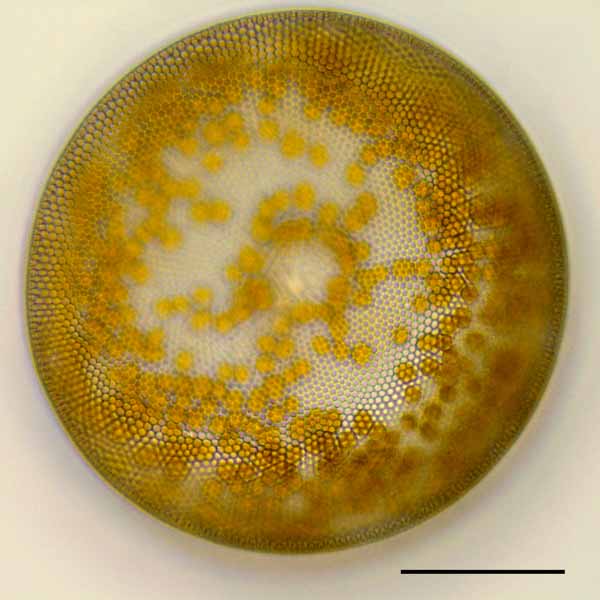
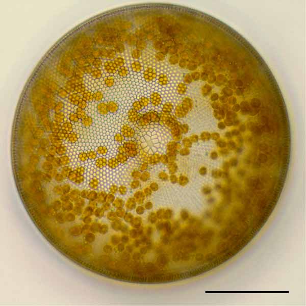
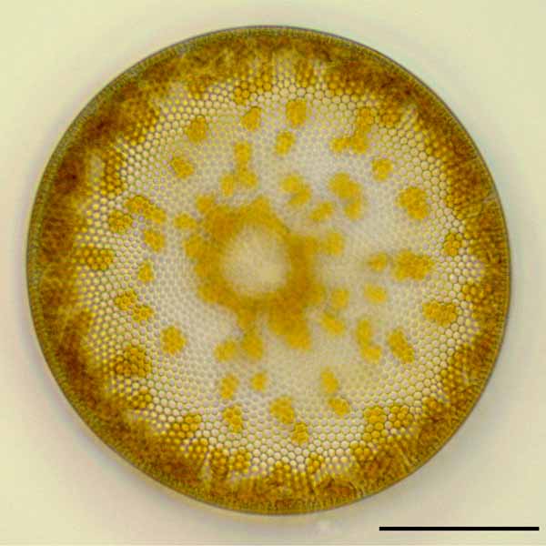
 写真04a〜写真04dはフォーカスを変えて撮影した画像です。
 大型の円盤状の細胞が単独で浮遊しています。
 殻には放射状に配列する胞紋列があります。
 円盤状の色素体が中心域の周りにリング状に集まっています。
写真05a
写真05b
20μm
20μm
コアミケイソウ (Coscinodiscus sp.)の画像
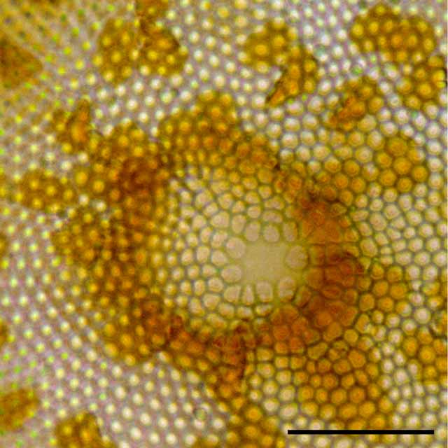
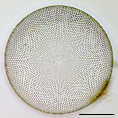
分類  :コアミケイソウ (Coscinodiscus sp.)
倍率  :1000倍 
採取  :海岸(茨城県 平磯海岸) 
 
時期  :2010年4月採取 表面海水温15.6℃ 


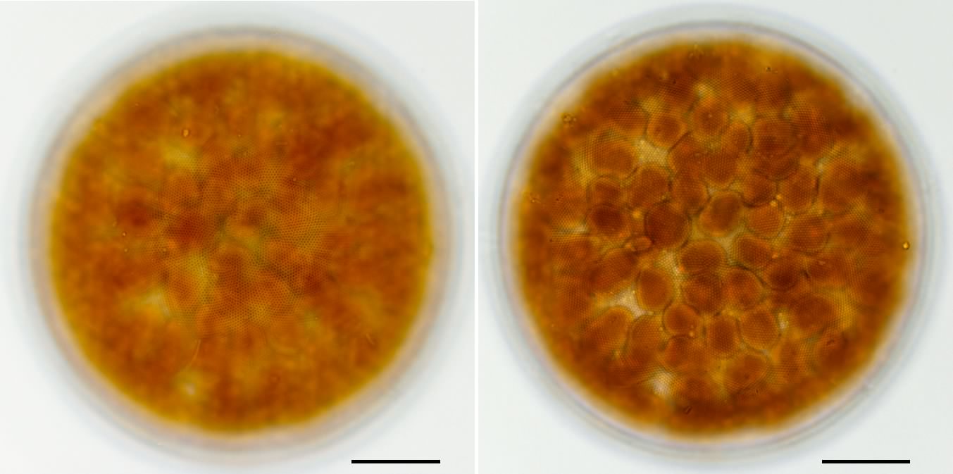
写真09a, 09b 
 被殻に焦点を合わせて撮影した画像です。
 写真01aは殻の全体像、写真01bは中心域の画像です。
 殻の表面(殻面)は平らではなく、中心域と周辺部で
 フォーカスの位置が異なります(写真01a)。
 殻には蜂の巣状の構造が(胞紋)があります(写真01b)。
 胞紋(areola)は、小さな孔状の構造です。
 胞紋は被殻の表面に垂直に開口しており、
 胞紋と胞紋との境界が蜂の巣状に見えています。

真09c, 09d 
 同じ珪藻の色素体にフォーカスを合わせた画像です。
 色素体は不規則な形態をしています。


写真09e, 09f
 
細胞内の核にフォーカスを合わせた画像です。
 核は細胞の中央からややずれた位置にあります。
 矢印の先に核があります。 
写真09f
写真09e
写真09d
写真09c
写真09b
写真09a
20μm
20μm
20μm
20μm
20μm
20μm
コアミケイソウ (Coscinodiscus sp.)の画像
分類  :コアミケイソウ (Coscinodiscus sp.)
倍率  :1000倍
 写真03a〜03dと同じ殻です。
20μm
20μm
20μm
20μm
写真03g
写真03h
写真03i
写真03j
コアミケイソウ (Coscinodiscus sp.)の画像
10μm
10μm
写真03e
写真03f
コアミケイソウ (Coscinodiscus sp.)の画像
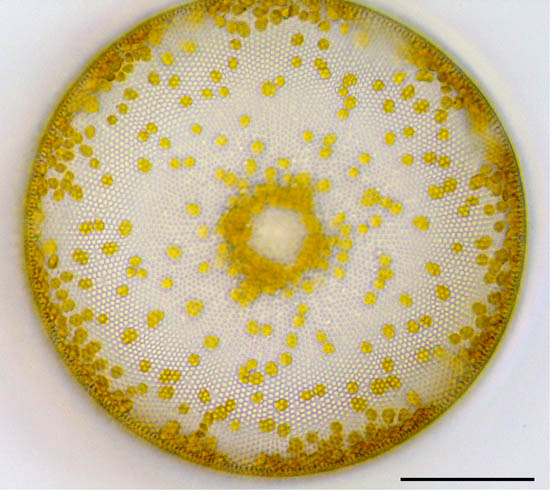
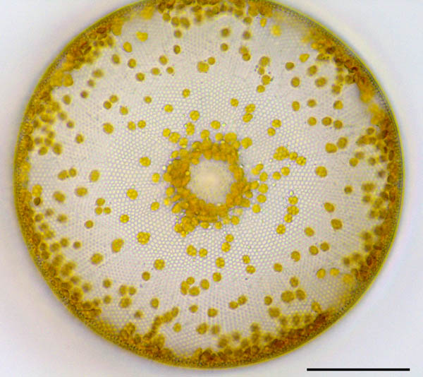
分類  :コアミケイソウ (Coscinodiscus sp.)
倍率  :1000倍
 写真02a〜02fと同じ細胞です。写真01の個体よりも色素体の数が多いように見えます。 
50μm
50μm
50μm
50μm
写真03a
写真03b
写真03c
写真03d
コアミケイソウ (Coscinodiscus sp.)の画像
20μm
20μm
20μm
20μm
写真02m
写真02n
写真02k
写真02l
コアミケイソウ (Coscinodiscus sp.)の画像
分類  :コアミケイソウ (Coscinodiscus sp.)
倍率  :1000倍
 写真02a〜02fと同じ細胞の中心付近を1000倍で観察しました。 
 殻に焦点が合っています。殻の中央には胞紋がなく、中央を取り囲む胞紋は周囲よりも大型です。
20μm
20μm
20μm
20μm
写真02g
写真02h
写真02i
写真02j
コアミケイソウ (Coscinodiscus sp.)の画像
写真01n
写真01m
分類  :コアミケイソウ (Coscinodiscus sp.)
倍率  :500倍 
採取  :海岸(東京都) 
 
時期  :2017年11月採取  

 東京湾のお台場で採取したコアミケイソウです。写真01a〜01fは焦点を変えながら撮影した画像です。 
 生きている細胞は殻表面の胞紋が不明瞭です。ただし、色素体の分布を明瞭に観察できます。
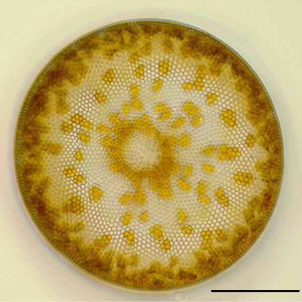
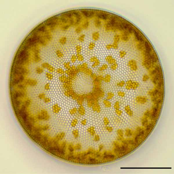
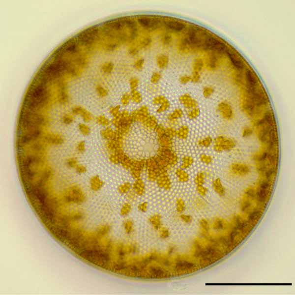
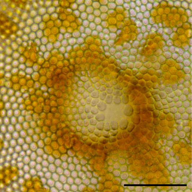
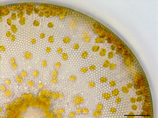
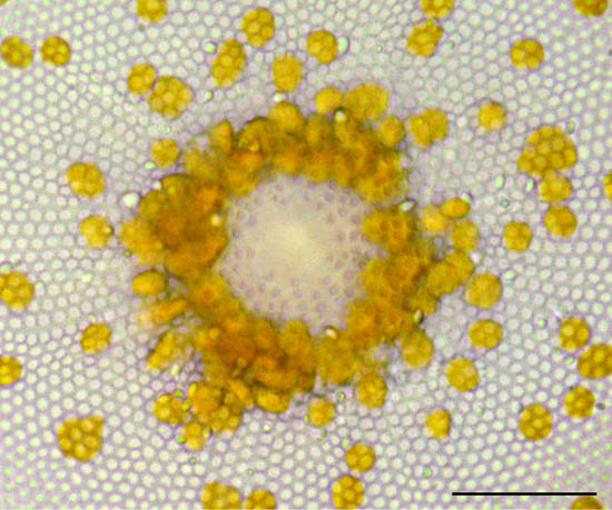
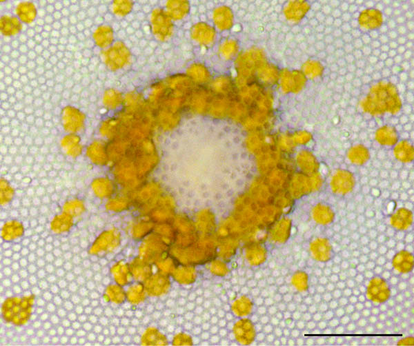
 コアミケイソウ (Coscinodiscus)は海洋の代表的な浮遊性珪藻で、プランクトンネットで採取できます。
 群体は作らず単独で浮遊しています。大型の珪藻で殻の構造(胞紋)も容易に観察できます。
 生きている細胞では殻内に多数の円盤状の色素体を見ることができます。
-------------------------------------------------------------------------------
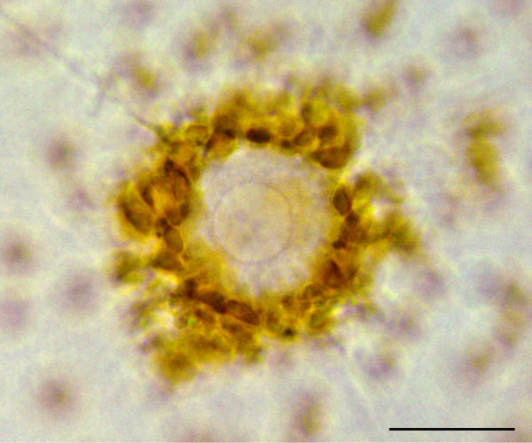
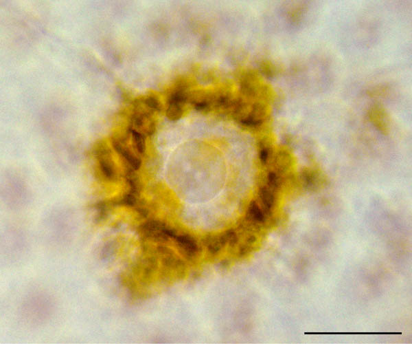
分類  :コアミケイソウ (Coscinodiscus sp.)
倍率  :1000倍
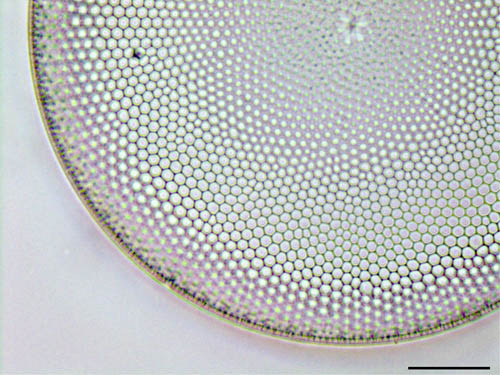
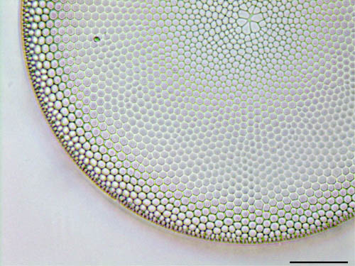
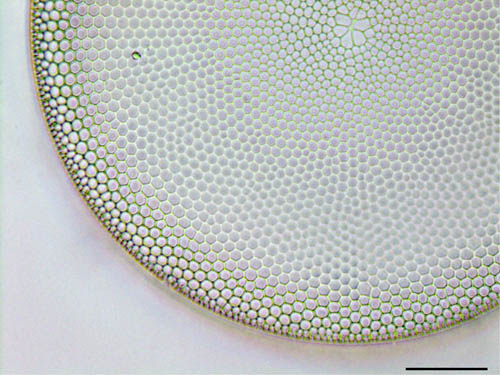
 写真03a〜03dと同じ殻の中心付近を1000倍で観察しました。 
 殻中央を取り囲む胞紋は周囲よりも大型です。
分類  :コアミケイソウ (Coscinodiscus sp.)
倍率  :1000倍
 写真01a〜01fと同じ細胞です。 
分類  :コアミケイソウ (Coscinodiscus sp.)
倍率  :1000倍
 写真01a〜01fと同じ細胞の中心付近を1000倍で観察しました。 
 写真01e〜01iは殻に焦点が合っています。殻の中央付近には胞紋がありません。
 写真01k〜01lは細胞の中心付近で、核と核に周囲に集まっている色素体を確認できます。
写真01h
コアミケイソウ (Coscinodiscus sp.)の画像
写真01g
50μm
写真02b
写真02a
写真01b
写真01a
最終更新日:2019/3/31
製作者:ねこのしっぽラボ
分類  :コアミケイソウ (Coscinodiscus sp.)
倍率  :500倍 
採取  :海岸(東京都) 
 
時期  :2017年11月採取  

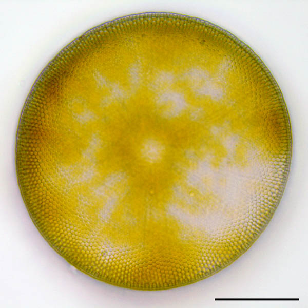
 東京湾のお台場で採取したコアミケイソウの殻です。原形質が綺麗になくなっています。 
 殻の模様(胞紋のパターン)は原形質がない方がよく見えます。
 殻中央の構造が写真02の種類と良く似ています。
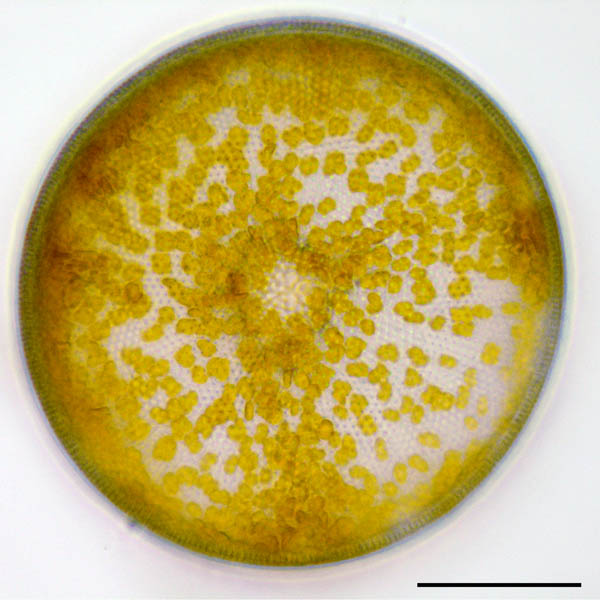
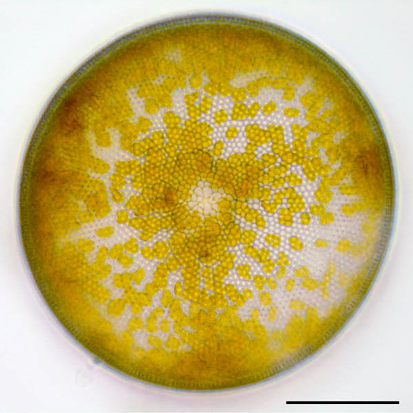
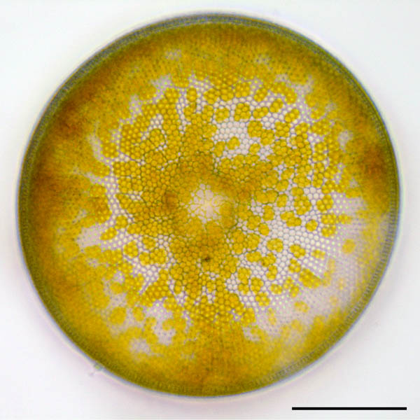
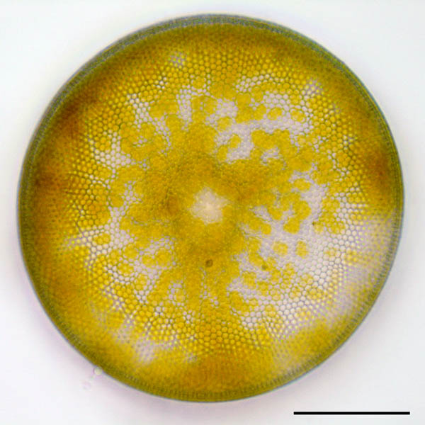
分類  :コアミケイソウ (Coscinodiscus sp.)
倍率  :500倍 
採取  :海岸(東京都) 
 
時期  :2017年11月採取  

 東京湾のお台場で採取したコアミケイソウです。写真02a〜02fは焦点を変えながら撮影した画像です。 
 写真01とl同時に採取しましたが、殻中央付近の胞紋の大きさが異なるので別種かもしれません。
コアミケイソウ (Coscinodiscus sp.)の画像
写真01p
写真01o
20μm
20μm
写真01j
コアミケイソウ (Coscinodiscus sp.)の画像
写真01i
20μm
20μm
50μm
50μm
50μm
50μm
50μm
50μm
写真02f
写真02e
写真01f
20μm
20μm
写真01e
写真01d
20μm
20μm
写真01l
コアミケイソウ (Coscinodiscus sp.)の画像
写真01k
20μm
20μm
写真01c
コアミケイソウ (Coscinodiscus sp.)の画像
トップページに戻ります。
50μm
50μm
50μm
50μm
50μm
写真02d
写真02c
コアミケイソウ (Coscinodiscus sp.)の画像